
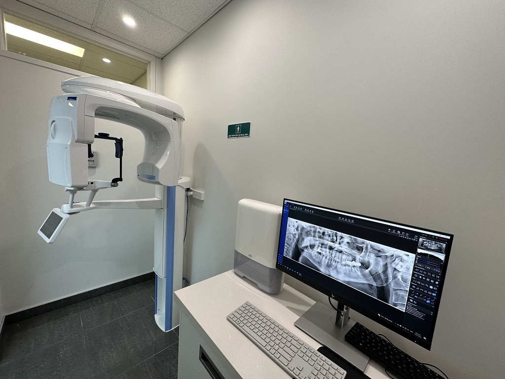
CONE-BEAM Computed Tomography (CBCT) Scan
Marina Dentists provide services for any dental pathology, restorative treatment, bite correction, surgical procedures like wisdom teeth extraction, as well as dental implantation using the latest technology that allows you to restore your teeth without additional operations, even in the presence of bone tissue atrophy.
To make an accurate diagnosis, monitor the progress and results of treatment, it is necessary to conduct high-quality diagnostics using a computed tomography (CT) scanner.
Marina Dentists have a new generation Planmeca CBCT scanner (Finland) for an in-depth study of the condition of all aspects of the oral cavity and jaw. The equipment allows for targeted X-rays of one tooth, a panoramic image of all teeth in the mouth (OPG or orthopantomogram), as well as 3D scanning.
Due to the minimum radiation exposure, the cone-beam computed tomography scanner is completely safe for the human body, even with frequent use.
The device is highly accurate, which is important for diagnosis - assessing the quality and quantity of bone tissue in preparation for placing the implant. Along with the CBCT scanner for 3D diagnostics, clinicians at Marina Dentists will use an intraoral scanner as well, which allows to obtain virtual impressions of the entire oral cavity and create a 3D virtual models required for implants, crowns ,bridges , dentures, or orthodontic treatment.Why you shouldn't be afraid to take X-Rays
To make an accurate diagnosis, monitor the progress and evaluate the results of treatment, it is important for this modern equipment to be used today. Many patients are concerned about how safe it is, as it may be necessary to take 2, 3, or sometimes more images.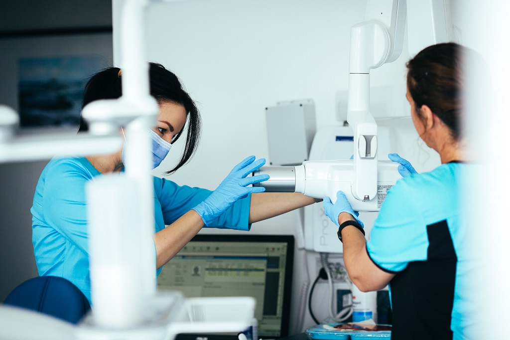
When X-rays, OPG and CT may be required
An X-ray is a sighting image of one or more teeth. An OPG or orthopantomogram is a panoramic image that captures both jaws. A CT scan is a computed tomography scan and captures three-dimensional images of the entire jaw. Each of these types of diagnostics are used for specific situations:- X-rays are performed for the treatment of a particular tooth: in the presence of caries (decay), pulpitis, periodontitis, suspected cysts, or granulomas. It allows you to determine the extent of tooth damage, as well as the condition of the tissues around the root,
- Orthopantomogram (OPG) is necessary for assessing periodontal structures, such as the jawbone. It is used for multiple dental treatments, such as orthodontic treatment or dental implants. It allows you to assess the condition and amount of bone tissue present, identify the position of the maxillary (nasal) sinuses of the upper jaw, nerves and other important anatomical elements.
- CT is performed for certain scenarios. Most often in the presence of tumors of the jaw (to determine their volume and dimensions), prior to dental implants placement, especially in difficult cases, wisdom teeth extraction , root canal treatment etc.
Is it dangerous to take X-Rays?
According to SanPiN1, when conducting X-ray procedures for prophylactic purposes, radiation exposure should not exceed 1000 microsieverts (μSv) per year . We are talking about prevention, since for therapeutic purposes the indicator can be much higher. In the number of shots, it looks like this:- 500 sighting shots (1-3 μSv),
- 80 OPG (13-17 μSv),
- 20 digital CT scans (50-60 μSv).
| In dentistry | In other fields of medicine | In life | Dangerous indicators |
| 1-3 μSv - one image | 30-60 μSv – one digital fluorography, 150-250 μSv – old-type FOG film | 5 μSv – 3 hours in front of a computer or TV | 750 thousand μSv - minor changes in blood composition |
| 13-17 μSv – one panoramic image (OPG) | 500-700 μSv – one mammogram procedure | 20-30 μSv – one 2-3-hour flight | 1 million μSv – mild radiation sickness |
| 50-60 μSv – one CT procedure in dentistry | 2000 μSv – one CT scan of the head | 2000-3000 μSv is the natural dose of radiation per person per year (food, solar radiation, air) | 7 million μSv is a lethal dosage of radiation |
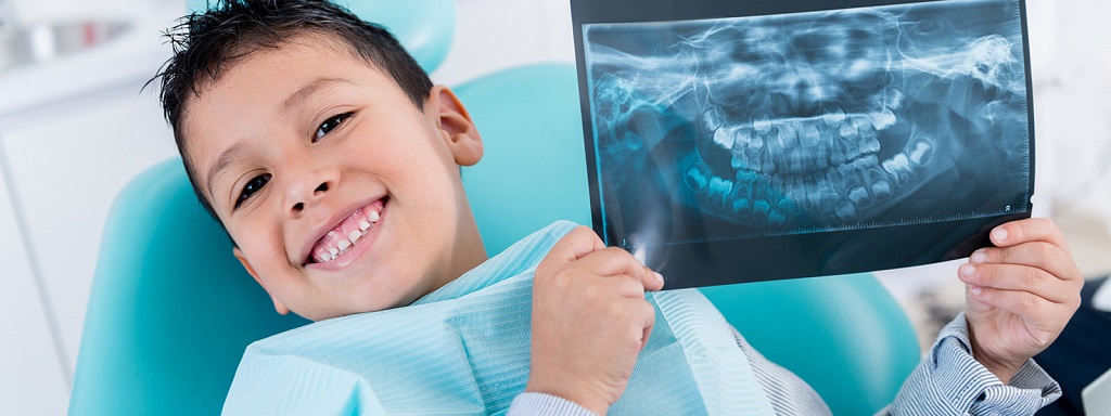
Is it possible to diagnose during pregnancy
According to the examinations, it is allowed to conduct X-ray examinations in the second half of pregnancy using protective equipment, provided that the radiation dose does not exceed the same 1000 μSv. However, it is recommended to refrain from carrying out X-rays in the first and last 12 weeks, i.e. in the first and last trimesters.Do not be afraid of diagnostic procedures during pregnancy. Even ordinary caries is an infection that, if left untreated, can spread throughout the body and lead to infection of the fetus. Therefore, it is better to get a small and completely safe dose of radiation than to carry out complex dental treatment blindly, not knowing how deep the decay or infection is.
 After the birth of the baby, i.e. during breastfeeding, radiography in dentistry can be carried out, (within reasonable limits). Radiation doses are minimal, so radiation does not accumulate in breast milk, there will be absolutely no harm to the child. Pumping and skipping feeding is also not required.